
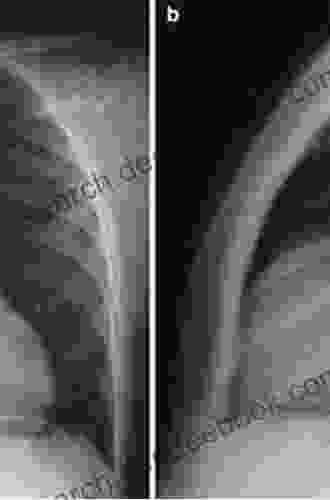
Pearls And Pitfalls In Thoracic Imaging

Thoracic imaging is a critical tool for diagnosing and managing a wide range of pulmonary and cardiovascular conditions. However, interpreting thoracic images can be challenging, and it is important to be aware of the potential pearls and pitfalls that may arise. In this article, we will discuss some of the key pearls and pitfalls in thoracic imaging, and provide tips on how to avoid them.
- Always correlate the images with the clinical history. The clinical history can provide valuable information about the patient's symptoms, risk factors, and previous medical history. This information can help to guide the interpretation of the images and ensure that the most appropriate diagnosis is made.
- Use a systematic approach to image interpretation. There are several different ways to approach thoracic image interpretation, but it is important to develop a systematic approach that ensures that all of the relevant findings are identified and interpreted. One common approach is to start by examining the lung parenchyma for any abnormalities, such as nodules, masses, or infiltrates. Next, the airways and mediastinum should be examined for any signs of obstruction or enlargement. Finally, the pleura and diaphragm should be examined for any abnormalities.
- Be aware of the normal variations in thoracic anatomy. The thoracic anatomy can vary significantly from person to person. It is important to be aware of these normal variations so that they are not mistaken for abnormalities. For example, the size and shape of the heart can vary depending on the patient's age, sex, and body habitus.
- Use appropriate imaging techniques. The choice of imaging technique will depend on the specific clinical question being asked. For example, chest radiography is a good screening tool for lung cancer, but it may not be sensitive enough to detect small nodules. Computed tomography (CT) is a more sensitive imaging technique that can be used to detect small nodules and other abnormalities in the lung parenchyma. Magnetic resonance imaging (MRI) is a good imaging technique for evaluating the mediastinum and great vessels.
- Over-interpreting the images. It is important to avoid over-interpreting the images and making a diagnosis that is not supported by the evidence. For example, a small nodule on chest radiography may not necessarily be a cancer. Further testing may be necessary to confirm the diagnosis.
- Missing subtle abnormalities. It is also important to avoid missing subtle abnormalities on the images. This can be difficult, especially if the abnormality is small or located in a complex area of the chest. It is important to carefully examine all of the images and to pay attention to any subtle changes that may be present.
- Being unaware of the limitations of the imaging technique. Each imaging technique has its own limitations. It is important to be aware of these limitations so that they are not misinterpreted as abnormalities. For example, chest radiography is not a good imaging technique for evaluating the mediastinum or great vessels.
- Not correlating the findings with the clinical history. It is important to correlate the findings on the images with the clinical history. This information can help to guide the interpretation of the images and ensure that the most appropriate diagnosis is made. For example, a patient with a history of smoking and a small nodule on chest radiography is at a higher risk of developing lung cancer than a patient with a negative smoking history.
Thoracic imaging is a valuable tool for diagnosing and managing a wide range of pulmonary and cardiovascular conditions. However, it is important to be aware of the potential pearls and pitfalls that may arise. By following the tips outlined in this article, you can avoid these pitfalls and ensure that you are interpreting thoracic images accurately and effectively.
- ACR Appropriateness Criteria for Thoracic Imaging
- RSNA Thoracic Imaging Learning Center
- American Thoracic Society Imaging Guidelines
5 out of 5
| Language | : | English |
| File size | : | 53514 KB |
| Text-to-Speech | : | Enabled |
| Screen Reader | : | Supported |
| Enhanced typesetting | : | Enabled |
| Print length | : | 234 pages |
5 out of 5
| Language | : | English |
| File size | : | 53514 KB |
| Text-to-Speech | : | Enabled |
| Screen Reader | : | Supported |
| Enhanced typesetting | : | Enabled |
| Print length | : | 234 pages |
Do you want to contribute by writing guest posts on this blog?
Please contact us and send us a resume of previous articles that you have written.
 Book
Book Novel
Novel Page
Page Chapter
 Text
Text Reader
 Library
 Bookmark
 Shelf
Shelf Glossary
 Bibliography
 Foreword
 Preface
 Synopsis
 Annotation
 Footnote
 Manuscript
 Scroll
 Tome
Tome Bestseller
 Library card
 Memoir
 Reference
 Encyclopedia
 Thesaurus
 Resolution
 Card Catalog
 Borrowing
 Stacks
 Archives
 Study
Study Scholarly
 Lending
 Reserve
 Reading Room
 Rare Books
 Literacy
 Dissertation
 Storytelling
 Theory
 Edmond J Keller
 Pierre Cormon
 Robert F Burgess
 Vanessa Riley
 Jennifer Cole Judd
 Shifio S Patterns
 Linda Lael Miller
 David Welch
 Jack Parker
 Virginia Mekkelson
 Divina Blackwell Bates
 Bill Kerber
 Scott Ritter
 Zbigniew Brzezinski
 Daniel W Drezner
 Jerry Sprout
 David M Addison
 Fiona Lucas
 David Freedberg
 Phoebe Morgan
Light bulbAdvertise smarter! Our strategic ad space ensures maximum exposure. Reserve your spot today!

 Floyd RichardsonWhat Lies Beneath: Sarah Rayne - An Enigmatic Tale of Love, Loss, and the...


 Joseph HellerThe Christmas Letters by Lee Smith: A Journey of Loss, Redemption, and the...
 Ralph Waldo EmersonHow Did That Get To My Table? Peanut Butter, Community Connections, and the...
 Jonathan FranzenFollow ·16k
 Liam WardFollow ·3.7k
 Jace MitchellFollow ·16.6k
 Jordan BlairFollow ·17.6k
 Wesley ReedFollow ·9.4k
 Jacob HayesFollow ·5.4k
 Donovan CarterFollow ·18.5k
 Isaac AsimovFollow ·2.3k

 Corbin Powell
My Little Bible Promises Thomas Nelson
In a world filled with uncertainty and...

 Tyler Nelson
Policing Rogue States: Open Media Series Explores Global...
In today's interconnected...

 Bret Mitchell
Musical Performance: A Comprehensive Guide to...
Immerse yourself in the...
 Juan Rulfo
Long Distance Motorcycling: The Endless Road and Its...
For many, the...

 Blake Kennedy
Vocal Repertoire for the Twenty-First Century: A...
The vocal repertoire of the twenty-first...
 Eric Hayes
One Hundred and Ninth on the Call Sheet! The Enigmatic...
In the vast panorama of Western films,...
5 out of 5
| Language | : | English |
| File size | : | 53514 KB |
| Text-to-Speech | : | Enabled |
| Screen Reader | : | Supported |
| Enhanced typesetting | : | Enabled |
| Print length | : | 234 pages |
