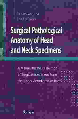
Manual for the Dissection of Surgical Specimens from the Upper Aerodigestive Tract: A Comprehensive Guide for Pathologists and Surgeons

The upper aerodigestive tract encompasses the anatomical structures involved in respiration, olfaction, speech, and deglutition, including the nasal cavity, paranasal sinuses, oral cavity, pharynx, and larynx. Surgical specimens from these regions are frequently encountered in pathology practice, and their proper dissection and examination are essential for accurate diagnosis and optimal patient management. This manual provides a comprehensive guide to the dissection of surgical specimens from the upper aerodigestive tract, tailored to the needs of both pathologists and surgeons.
Nasal Cavity and Paranasal Sinuses
Specimen Collection and Handling
Specimens of the nasal cavity and paranasal sinuses are typically obtained through endoscopic or open surgical approaches. Fresh specimens should be immediately placed in a preservative solution, such as formalin or Bouin's solution, to prevent autolysis and allow for proper fixation.
5 out of 5
| Language | : | English |
| File size | : | 28875 KB |
| Text-to-Speech | : | Enabled |
| Screen Reader | : | Supported |
| Enhanced typesetting | : | Enabled |
| Print length | : | 159 pages |
Dissection Technique
1. External Examination: Examine the specimen for any external abnormalities, such as polyps, ulcerations, or masses. Note their size, location, and relation to surrounding structures. 2. Incision: Create a vertical incision along the midline of the nasal septum to separate the nasal cavities. Extend the incision posteriorly to the nasopharynx. 3. Turbinate Examination: Remove the inferior and middle turbinates to expose the lateral nasal wall. Inspect the mucosa for any abnormalities, such as hypertrophy, atrophy, or ulceration. 4. Ethmoid and Sphenoid Sinuses: Remove the uncinate process to expose the ethmoid labyrinth. Carefully dissect the ethmoid bullae and cells to visualize the sphenoid sinus. Examine the mucosa and bony walls for any abnormalities.
Histological Interpretation
The histological examination of nasal and paranasal sinus specimens focuses on assessing the mucosal lining, underlying stroma, and bony walls. Common pathological findings include:
* Chronic rhinitis: Characterized by mucosal inflammation, edema, and hyperplasia. * Polyp formation: Proliferations of edematous mucosal tissue projecting into the nasal cavity. * Sinusitis: Inflammation of the paranasal sinus mucosa, with thickening, edema, and neutrophil infiltration. * Neoplasms: Various types of benign and malignant tumors can arise in the nasal cavity and paranasal sinuses, including squamous cell carcinoma, adenocarcinoma, and lymphomas.
Oral Cavity
Specimen Collection and Handling
Surgical specimens from the oral cavity may include biopsies, resections, and radical or partial glossectomy specimens. Fresh specimens should be immediately placed in formalin or Bouin's solution.
Dissection Technique
1. External Examination: Examine the specimen for any external abnormalities, such as ulcers, masses, or leukoplakia. Note their location and relation to adjacent structures. 2. Incision: If the specimen includes the tongue or floor of mouth, make a midline sagittal incision to expose the underlying structures. 3. Mucosal Examination: Inspect the oral mucosa for any abnormalities, such as hyperplasia, atrophy, or ulceration. Palpate the submucosa to assess for any induration or masses. 4. Tongue and Floor of Mouth: Examine the tongue muscle, mucosa, and associated lymph nodes. Inspect the floor of mouth for any abnormalities, such as ranulas or sialoliths.
Histological Interpretation
The histological examination of oral cavity specimens focuses on assessing the mucosal lining, underlying stroma, and associated structures. Common pathological findings include:
* Leukoplakia: A premalignant condition characterized by white, thickened, and adherent mucosal plaques. * Dysplasia: Abnormal cellular changes in the mucosa, ranging from mild to severe, indicating a risk of malignant transformation. * Squamous cell carcinoma: The most common malignancy of the oral cavity, with variable histological features, including keratinization, dyskeratosis, and cellular atypia. * Salivary gland tumors: A diverse group of benign and malignant neoplasms arising from the salivary glands, requiring specific immunohistochemical markers for diagnosis.
Pharynx
Specimen Collection and Handling
Surgical specimens from the pharynx may include biopsies, resections, or radical neck dissection specimens. Fresh specimens should be immediately placed in formalin or Bouin's solution.
Dissection Technique
1. External Examination: Examine the specimen for any external abnormalities, such as masses, ulcerations, or enlarged lymph nodes. Note their location and relation to surrounding structures. 2. Incision: If the specimen includes the oropharynx, make a midline sagittal incision to expose the underlying structures. Extend the incision posteriorly to the nasopharynx and anteriorly to the hypopharynx. 3. Mucosal Examination: Inspect the pharyngeal mucosa for any abnormalities, such as hyperplasia, atrophy, or ulceration. Palpate the submucosa to assess for any induration or masses. 4. Tonsil Examination: Remove the tonsils and examine them for any lymphoid hyperplasia, crypts, or abscesses.
Histological Interpretation
The histological examination of pharyngeal specimens focuses on assessing the mucosal lining, underlying stroma, and associated lymphoid tissue. Common pathological findings include:
* Chronic pharyngitis: Characterized by mucosal inflammation, edema, and hyperplasia. * Tonsillitis: Inflammation of the tonsils, with lymphoid hyperplasia, follicular formation, and neutrophil infiltration. * Squamous cell carcinoma: The most common malignancy of the pharynx, with variable histological features, including keratinization, dyskeratosis, and cellular atypia. * Nasopharyngeal carcinoma: A distinct type of carcinoma arising from the nasopharyngeal mucosa, often associated with Epstein-Barr virus infection and exhibiting a characteristic histopathology.
Larynx
Specimen Collection and Handling
Surgical specimens from the larynx may include biopsies, laryngectomy specimens, or radical neck dissection specimens. Fresh specimens should be immediately placed in formalin or Bouin's solution.
Dissection Technique
1. External Examination: Examine the specimen for any external abnormalities, such as masses, ulcerations, or enlarged lymph nodes. Note their location and relation to surrounding structures. 2. Incision: If the specimen includes the larynx, make a vertical incision through the thyroid cartilage to expose the laryngeal lumen. Extend the incision superiorly to the epiglottis and inferiorly to the trachea. 3. Mucosal Examination: Inspect the laryngeal mucosa for any abnormalities, such as hyperplasia, atrophy, or ulceration. Palpate the submucosa to assess for any induration or masses. 4. Vocal Cord Examination: Examine the vocal cords for any abnormalities, such as nodules, polyps, or granulomas. Assess their mobility and symmetry.
Histological Interpretation
The histological examination of laryngeal specimens focuses on assessing the mucosal lining, underlying stroma, and associated cartilaginous structures. Common pathological findings include:
* Chronic laryngitis: Characterized by mucosal inflammation, edema, and hyperplasia. * Papillomatosis: Warty growths on the laryngeal mucosa, often caused by human papillomavirus infection. * Squamous cell carcinoma: The most common malignancy of the larynx, with variable histological features, including keratinization, dyskeratosis, and cellular atypia. * Vocal cord lesions: A variety of benign and malignant lesions can affect the vocal cords, including nodules, polyps, and granulomas.
This manual provides a comprehensive guide to the dissection of surgical specimens from the upper aerodigestive tract, covering the nasal cavity and paranasal sinuses, oral cavity, pharynx, and larynx. By following the described techniques, pathologists and surgeons can ensure optimal specimen preparation and histological evaluation, facilitating accurate diagnosis and appropriate patient management.
5 out of 5
| Language | : | English |
| File size | : | 28875 KB |
| Text-to-Speech | : | Enabled |
| Screen Reader | : | Supported |
| Enhanced typesetting | : | Enabled |
| Print length | : | 159 pages |
Do you want to contribute by writing guest posts on this blog?
Please contact us and send us a resume of previous articles that you have written.
 Novel
Novel Chapter
 Story
Story Reader
 E-book
 Magazine
 Newspaper
 Bookmark
 Shelf
Shelf Glossary
 Foreword
 Annotation
 Manuscript
 Tome
Tome Bestseller
 Classics
 Library card
 Biography
 Autobiography
 Memoir
 Reference
 Encyclopedia
 Dictionary
 Thesaurus
 Character
 Librarian
 Catalog
 Stacks
 Archives
 Periodicals
 Research
 Scholarly
 Lending
 Reserve
 Journals
 Rare Books
 Interlibrary
 Storytelling
 Reading List
 Theory
 I L Willams
 William M Tsutsui
 Johnny Cox
 Brian Wolf
 Brian Curtis
 Robert Coles
 Delores Henriques
 James Arena
 Carmen Rita
 Timothy Beatley
 Tzeli Hadjidimitriou
 Rachmi Diyah Larasati
 Karen Jones
 Guilherme Douglas Balista
 Tara Benham
 Miles Davis
 Timothy Garton Ash
 Libby Worth
 John William Polidori
 Matilda Walsh
Light bulbAdvertise smarter! Our strategic ad space ensures maximum exposure. Reserve your spot today!
 Hunter MitchellFollow ·6.2k
 James HayesFollow ·15.5k
 Dalton FosterFollow ·13.7k
 Colt SimmonsFollow ·2.2k
 Osamu DazaiFollow ·14.4k
 W.H. AudenFollow ·9.6k
 Ivan TurgenevFollow ·16.8k
 José MartíFollow ·19.6k

 Corbin Powell
My Little Bible Promises Thomas Nelson
In a world filled with uncertainty and...

 Tyler Nelson
Policing Rogue States: Open Media Series Explores Global...
In today's interconnected...

 Bret Mitchell
Musical Performance: A Comprehensive Guide to...
Immerse yourself in the...

 Juan Rulfo
Long Distance Motorcycling: The Endless Road and Its...
For many, the...

 Blake Kennedy
Vocal Repertoire for the Twenty-First Century: A...
The vocal repertoire of the twenty-first...

 Eric Hayes
One Hundred and Ninth on the Call Sheet! The Enigmatic...
In the vast panorama of Western films,...
5 out of 5
| Language | : | English |
| File size | : | 28875 KB |
| Text-to-Speech | : | Enabled |
| Screen Reader | : | Supported |
| Enhanced typesetting | : | Enabled |
| Print length | : | 159 pages |


