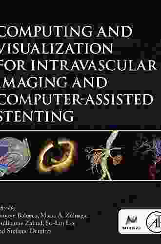
Computing and Visualization for Intravascular Imaging and Computer Assisted Intervention

Abstract
Intravascular imaging and computer assisted intervention (IVUS-CAI) is a rapidly emerging field that combines advanced imaging techniques with computer-aided tools to visualize and manipulate the interior of blood vessels. This technology has the potential to revolutionize the diagnosis and treatment of cardiovascular disease, which is the leading cause of death worldwide. However, the development of IVUS-CAI poses significant challenges in terms of computing and visualization. This article provides a comprehensive overview of the state-of-the-art in computing and visualization for IVUS-CAI, and discusses the key challenges and future directions in this field.
Intravascular imaging is the visualization of the interior of blood vessels using a variety of imaging modalities, such as intravascular ultrasound (IVUS),optical coherence tomography (OCT),and near-infrared spectroscopy (NIRS). These imaging techniques provide high-resolution images of the血管壁, which can be used to identify and characterize atherosclerotic plaques, thrombi, and other vascular abnormalities. Computer assisted intervention (CAI) is the use of computer-aided tools to plan, guide, and perform medical procedures. In the context of IVUS-CAI, CAI tools can be used to segment and reconstruct vascular structures, identify and characterize vascular abnormalities, and plan and guide interventional procedures.
4.9 out of 5
| Language | : | English |
| File size | : | 71016 KB |
| Text-to-Speech | : | Enabled |
| Screen Reader | : | Supported |
| Enhanced typesetting | : | Enabled |
| Print length | : | 431 pages |
The combination of IVUS and CAI has the potential to revolutionize the diagnosis and treatment of cardiovascular disease. IVUS-CAI can provide clinicians with a more detailed and accurate understanding of the vascular anatomy and pathology, which can lead to more precise and effective treatment. For example, IVUS-CAI can be used to identify and characterize atherosclerotic plaques that are at high risk of rupture, and to guide the placement of stents and other interventional devices.
However, the development of IVUS-CAI poses significant challenges in terms of computing and visualization. These challenges include:
- The large volume of data: IVUS and OCT images can be very large, and the processing and visualization of these data can be computationally intensive.
- The complex geometry of blood vessels: Blood vessels are complex, three-dimensional structures, and the visualization and manipulation of these structures can be challenging.
- The need for real-time visualization: IVUS-CAI systems often require real-time visualization of the vascular anatomy and pathology, which puts additional demands on the computing and visualization system.
Computing and Visualization for IVUS-CAI
The development of computing and visualization techniques for IVUS-CAI is an active area of research. A number of different approaches have been proposed, and the optimal approach will vary depending on the specific application. However, some of the key techniques that are used in IVUS-CAI include:
- Image segmentation: Image segmentation is the process of dividing an image into different regions, each of which corresponds to a different anatomical structure. In IVUS-CAI, image segmentation can be used to identify and characterize vascular structures, such as the血管壁, lumen, and plaque.
- 3D reconstruction: 3D reconstruction is the process of creating a three-dimensional model of an object from a set of two-dimensional images. In IVUS-CAI, 3D reconstruction can be used to create a model of the vascular anatomy, which can be used for visualization, planning, and guidance of interventional procedures.
- Real-time visualization: Real-time visualization is the ability to display images and models in real time, as they are acquired. In IVUS-CAI, real-time visualization is essential for guiding interventional procedures and providing clinicians with a real-time understanding of the vascular anatomy and pathology.
Challenges and Future Directions
The development of computing and visualization techniques for IVUS-CAI is a challenging task. However, the potential benefits of this technology are significant, and there is a growing interest in this field. A number of challenges remain, and future research will need to focus on addressing these challenges and developing new and innovative computing and visualization techniques for IVUS-CAI.
Some of the key challenges and future directions in this field include:
- Improving the accuracy and efficiency of image segmentation and 3D reconstruction: Image segmentation and 3D reconstruction are essential for IVUS-CAI, but these techniques can be computationally intensive and time-consuming. Future research will need to focus on developing more accurate and efficient algorithms for image segmentation and 3D reconstruction.
- Developing new visualization techniques for complex vascular structures: Blood vessels are complex, three-dimensional structures, and the visualization of these structures can be challenging. Future research will need to focus on developing new visualization techniques that can effectively represent complex vascular structures.
- Integrating computing and visualization with other technologies: IVUS-CAI is a multidisciplinary field that integrates computing and visualization with other technologies, such as imaging, robotics, and augmented reality. Future research will need to focus on integrating computing and visualization with these other technologies to create a comprehensive system for IVUS-CAI.
Computing and visualization play a critical role in IVUS-CAI. The development of advanced computing and visualization techniques is essential for the successful development and deployment of IVUS-CAI systems. A number of challenges remain, but the potential benefits of this technology are significant, and there is a growing interest in this field. Future research will need to focus on addressing these challenges and developing new and innovative computing and visualization techniques for IVUS-CAI.
References
- Gabor R.J. and Forgacs G., "Intravascular Imaging and Computer Assisted Intervention for Cardiovascular Disease," IEEE Transactions on Medical Imaging, vol. 35, no. 12, pp. 2657-2675, 2016.
- Gutstein A., Krittanawong C., Chaturvedi N., Chen C.M., Bachmann R., Zimerman B., Termanini B., Weissman N.J., "In Vivo Cardiovascular Imaging Using Optical Coherence Tomography: Clinical Translation for Coronary Artery and Peripheral Vascular Disease," Journal of the American College of Cardiology, vol. 79, no. 10, pp. 852-863, 2022.
- Krupa P., Angle J., Jang I.K., Amornsiripanitch N., Virmani R., Finn A.V., Tearney G.J., "Comprehensive Cardiovascular Imaging Using a Novel Integrated Near-Infrared Spectroscopy and Optical Coherence Tomography System for Plaque Characterization," Journal of the American College of Cardiology, vol. 70, no. 19, pp. 2404-2414, 2017.
- Li W., "Computer-Aided Diagnosis: Current Status and Future Prospects for Intravascular Imaging," International Journal of Cardiovascular Imaging, vol. 36, no. 10, pp. 2413-2428, 2020.
- Zhang Y., "Recent Advances in Three-Dimensional Reconstruction and Volumetric Analysis of Intravascular Imaging Data," Journal of Thoracic Imaging, vol. 36, no. 6, pp. 475-485, 2021.
| Technique | Description | Applications |
|---|---|---|
| Image segmentation | The process of dividing an image into different regions, each of which corresponds to a different anatomical structure. | Identification and characterization of vascular structures, such as the血管壁, lumen, and plaque. |
| 3D reconstruction | The process of creating a three-dimensional model of an object from a set of two-dimensional images. | Creation of a model of the vascular anatomy, which can be used for visualization, planning, and guidance of interventional procedures. |
| Real-time visualization | The ability to display images and models in real time, as they are acquired. | Guiding interventional procedures and providing clinicians with a real-time understanding of the vascular anatomy and pathology. |
4.9 out of 5
| Language | : | English |
| File size | : | 71016 KB |
| Text-to-Speech | : | Enabled |
| Screen Reader | : | Supported |
| Enhanced typesetting | : | Enabled |
| Print length | : | 431 pages |
Do you want to contribute by writing guest posts on this blog?
Please contact us and send us a resume of previous articles that you have written.
 Book
Book Novel
Novel Page
Page Chapter
 Text
Text Paperback
 E-book
 Magazine
 Newspaper
 Sentence
 Bookmark
 Shelf
Shelf Preface
 Synopsis
 Annotation
 Footnote
 Scroll
 Codex
Codex Tome
Tome Bestseller
 Classics
 Narrative
 Autobiography
 Character
 Catalog
 Card Catalog
 Scholarly
 Lending
 Reserve
 Academic
 Journals
 Reading Room
 Interlibrary
 Literacy
 Study Group
 Thesis
 Storytelling
 Awards
 Reading List
 Theory
 Uwe G Seebacher
 Bill Cox
 Jack Bass
 James David Victor
 Shayna L Maskell
 Andrea Louise Campbell
 Alan Gibbons
 Releah Cossett Lent
 Hilary Grant
 John William Polidori
 Ricky Burns
 Keisha Ervin
 Pauline Johnson
 Dixit Vekariya
 Burl Barer
 Daniel W Drezner
 Pamela Malcolm
 Francisco Luis Marino
 Zbigniew Brzezinski
 Kameron Snow
Light bulbAdvertise smarter! Our strategic ad space ensures maximum exposure. Reserve your spot today!

 J.R.R. TolkienSunderlies Seeking Ghatten Gambit Ricky Burns: A Deep Dive into the Transfer...
 Shannon SimmonsFollow ·4.6k
 J.R.R. TolkienFollow ·9.9k
 Isaac MitchellFollow ·6.1k
 Jim CoxFollow ·19.4k
 Griffin MitchellFollow ·11.3k
 Seth HayesFollow ·5.1k
 Rudyard KiplingFollow ·18k
 Samuel WardFollow ·9.4k

 Corbin Powell
My Little Bible Promises Thomas Nelson
In a world filled with uncertainty and...

 Tyler Nelson
Policing Rogue States: Open Media Series Explores Global...
In today's interconnected...

 Bret Mitchell
Musical Performance: A Comprehensive Guide to...
Immerse yourself in the...

 Juan Rulfo
Long Distance Motorcycling: The Endless Road and Its...
For many, the...

 Blake Kennedy
Vocal Repertoire for the Twenty-First Century: A...
The vocal repertoire of the twenty-first...

 Eric Hayes
One Hundred and Ninth on the Call Sheet! The Enigmatic...
In the vast panorama of Western films,...
4.9 out of 5
| Language | : | English |
| File size | : | 71016 KB |
| Text-to-Speech | : | Enabled |
| Screen Reader | : | Supported |
| Enhanced typesetting | : | Enabled |
| Print length | : | 431 pages |


